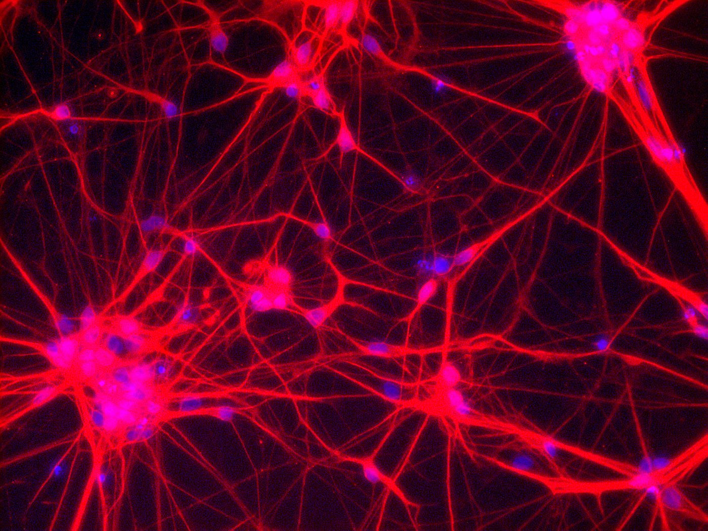
Neurons – Network of neuronal cells (Blue: nucleus; Red: neurons) [Copyright: Institute for Transfusion Medicine, UK Essen]
![[Translate to En:]](/fileadmin/_processed_/0/7/csm_02_Organoid_d156a1bd42.jpg "[Translate to En:]")
[Translate to En:]
Scientists have been able to generate brain-like 3D structures from stem cells, often referred to as "mini-brains" or “brain in a dish”, in the laboratory for several years. These organoids, which are 2-4 mm in size, are a very valuable model system, especially in developmental biology, but of course they do not possess all the properties of a human brain yet. Now, for the first time, several scientists from the Stem Cell Network NRW, together with other scientists, have succeeded in producing brain organoids that already have primitive sensory structures, i.e. the brain organoids can react to light stimuli. These novel models will help scientists to better investigate and understand the complex structures between the brain and the optic nerve in future. The results were published in the renowned journal Cell Stem Cell: Gabriel et al.: "Human brain organoids assemble functionally integrated bilateral optic vesicles", August 17, 2021.
The researchers' starting material are induced pluripotent stem cells (iPSC), from which they can generate human brain organoids in the laboratory using a specific protocol. Through specific adaptations in the protocol, the researchers have now succeeded in having the brain organoids form optic nerves on both sides of the forebrain. After about 30 days in the petri dish, the formation of optic vesicles could be observed on the organoids, and within 60 days they developed further into visible structures. The brain organoids with optic nerves are called "OVB organoids" (optic vesicle-containing brain organoids) and have structural similarities to the human eye: primitive corneal epithelial and lens-like cells, retinal pigment epithelia, retinal progenitor cells, axon-like projections, and electrically active neuronal networks (a kind of retina), which react to light incidence and transmit the signals to the interior of the approx. 2 mm miniature brains.
So far it has not been possible to demonstrate interactions between different areas (organs) within a single organoid. In the present study, the scientists were able for the first time to demonstrate such interactions using "photobleaching" experiments: The brain organoids were able to organise primitive sensory structures in the forebrain to a certain extent. The novel models of human brain organoids with bilaterally symmetrical optic nerves open up significant possibilities to better understand the complex development of the brain in the future and also make an important contribution in the study of diseases. “These organoids can help to study brain-eye interactions during embryo development, model congenital retinal disorders, and generate patient-specific retinal cell types for personalized drug testing and transplantation therapies.” outlines Prof. Jay Gopalakrishnan (University Hospital Düsseldorf), who is the last author of the publication.
Prof. Gopalakrishnan will present the results of this study in our Spotlight Series on Thursday, 30.09.2021. Participation is free of charge. If you are interested, please contact info[at]stammzellen.nrw.de
Research profiles of the participating working groups from the Stem Cell Network NRW:
c/o Life Science Center
Merowingerplatz 1
D 40225 Düsseldorf
Funded by:
![]()